
Glaucoma is the silent thief of sight - it often progresses without pain, and many people don’t realize they have it until noticeable vision loss occurs. The good news? A simple, routine glaucoma eye exams can catch the disease early, preserving vision and quality of life. This article explains why keeping up with regular eye check‑ups is non‑negotiable if you want to stay ahead of glaucoma.
Key Takeaways
- Glaucoma damage is irreversible, but early detection through regular exams can stop progression.
- Intraocular pressure, optic nerve imaging, and visual‑field tests are the three main screening tools.
- People over 40, those with family history, or certain medical conditions should schedule exams at least annually.
- Open‑angle and angle‑closure glaucoma require different management strategies.
- Collaboration between optometrists and ophthalmologists ensures comprehensive care.
Understanding Glaucoma
Glaucoma is a group of eye diseases that damage the optic nerve, often because of elevated intraocular pressure. It’s the second leading cause of blindness worldwide, affecting over 76 million people. The disease progresses slowly, and many individuals experience no symptoms until peripheral vision is already lost. Knowing the mechanics of glaucoma helps you appreciate why routine monitoring is essential.
Why Regular Eye Exams Matter
A regular eye exam is a comprehensive check‑up performed by an eye‑care professional that assesses visual acuity, eye pressure, optic nerve health, and more. These appointments are more than a vision‑sharpness test - they’re a window into the health of the optic nerve and the fluid dynamics inside the eye.
Because glaucoma can develop without obvious signs, doctors rely on three objective measurements during each visit:
- Intraocular pressure (IOP) reading
- Optic nerve head evaluation
- Visual‑field (perimetry) testing
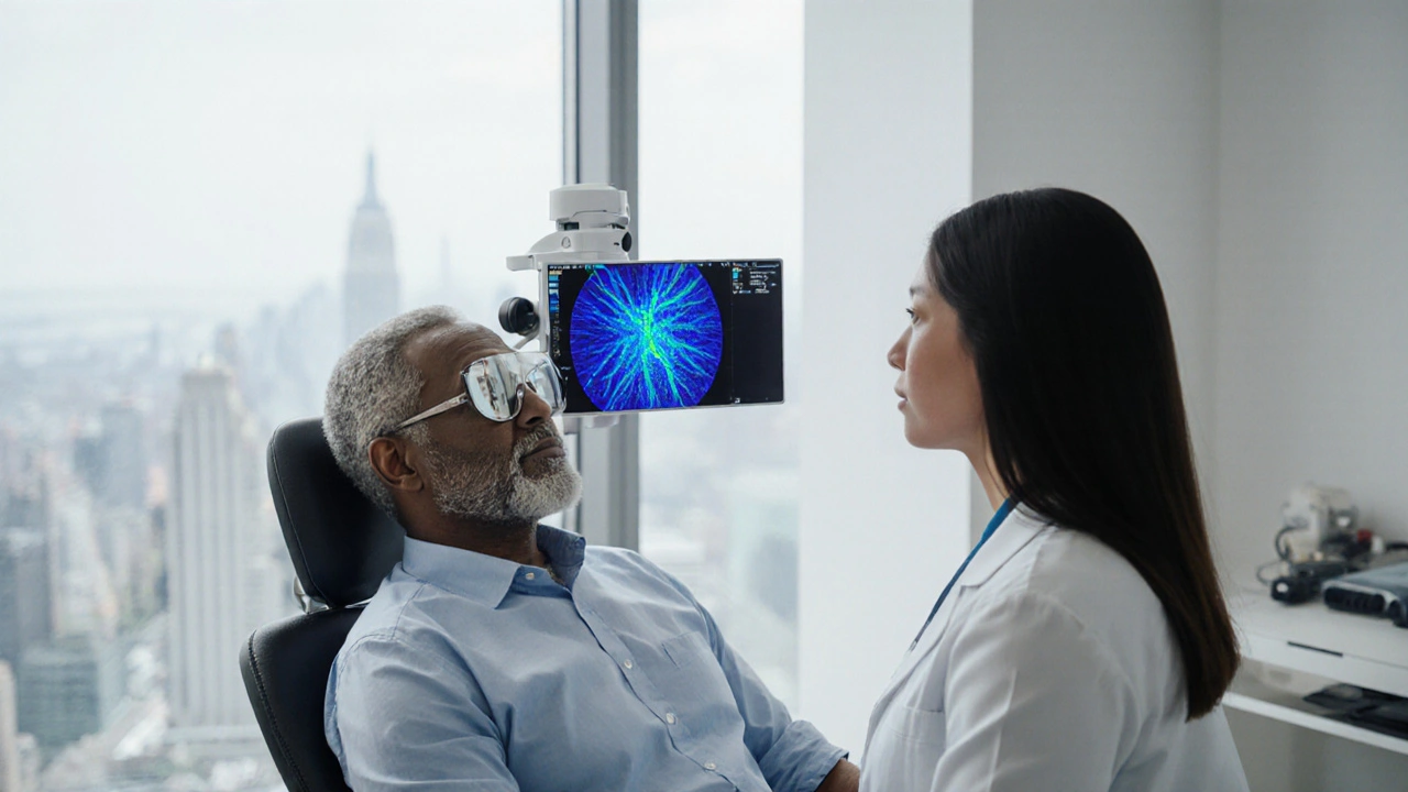
How Eye Exams Detect Glaucoma
Intraocular pressure is the fluid pressure inside the eye, measured in millimeters of mercury (mmHg). Elevated IOP is a major risk factor, and tonometry devices give a quick, painless reading. Normal ranges sit between 10‑21 mmHg; sustained readings above 22 mmHg warrant closer investigation.
Next, clinicians examine the optic nerve using ophthalmoscopy or advanced imaging such as OCT (optical coherence tomography). The optic disc’s cupping - a hollowing that appears as a dark center - can indicate nerve fiber loss even when pressure looks normal.
Finally, visual‑field testing maps a patient’s peripheral vision. The test reveals blind spots that are characteristic of early glaucoma, often before the patient notices any trouble reading or navigating.
Who Should Get Checked and How Often?
Risk factors are a powerful predictor. If you fit any of the following, schedule an exam at least once a year:
- Age 40+ (risk rises sharply after 60)
- Family history of glaucoma
- High myopia (nearsightedness)
- Diabetes or hypertension
- African, Hispanic, or Asian ancestry (certain types are more prevalent)
People with borderline IOP or early optic‑nerve changes may need more frequent monitoring, sometimes every six months. Your eye‑care professional will tailor the schedule based on your individual risk profile.
Types of Glaucoma: A Quick Comparison
| Feature | Open‑Angle Glaucoma | Angle‑Closure Glaucoma |
|---|---|---|
| Onset | Gradual, often asymptomatic | Sudden, can cause severe eye pain |
| Cause | Drainage canals work but fluid builds up | Anterior chamber angle closes, blocking drainage |
| Prevalence | ~90% of cases worldwide | ~10% of cases, more common in Asian populations |
| Treatment | Eye‑drops, laser trabeculoplasty, surgery | Laser iridotomy, medication, possible surgery |
Managing Glaucoma After Detection
Once glaucoma is confirmed, a multidisciplinary approach keeps the disease in check.
Ophthalmologist is a medical doctor specialized in eye diseases who can prescribe treatments and perform surgeries. They often work with optometrist is a primary eye‑care provider who conducts routine exams, detects early signs, and refers patients as needed. Together they decide on the best regimen, which may include:
- Medicated eye‑drops: Prostaglandin analogs (e.g., latanoprost) lower IOP by increasing fluid outflow.
- Laser therapy: Selective laser trabeculoplasty (SLT) improves drainage without incisions.
- Surgical options: Trabeculectomy or minimally invasive glaucoma surgery (MIGS) create new pathways for fluid.
Adherence matters. Studies show that up to 40% of patients miss doses, leading to faster progression. Setting reminders, using drop‑caddies, and regular follow‑up visits dramatically improve outcomes.
Common Pitfalls and Pro Tips
Even with the best care plan, some habits sabotage success:
- Skipping exams: Missing an annual check‑up can let pressure spikes go unnoticed.
- Self‑diagnosing: Online symptom checkers aren’t a substitute for tonometry and optic‑nerve imaging.
- Ignoring side effects: Some drops cause redness or eyelash growth; talk to your doctor before stopping them.
Here are three quick actions to keep your eyes safe:
- Mark your calendar for the next eye exam right after leaving the office.
- Keep a log of drop usage and any side effects.
- Ask your eye‑care professional about lifestyle factors - regular exercise, a balanced diet, and limiting caffeine can modestly support eye‑pressure control.

Frequently Asked Questions
Can glaucoma be cured?
No. Glaucoma damage to the optic nerve is permanent. However, early detection and treatment can halt or slow further loss, preserving the vision you still have.
How often should a healthy adult get an eye exam for glaucoma?
If you have no risk factors, a comprehensive exam every two years is sufficient. Anyone with risk factors should be examined annually or as advised by their eye‑care professional.
Are eye‑drops the only treatment?
Eye‑drops are first‑line, but laser therapy and surgery are effective alternatives or adjuncts when drops don’t achieve target pressure.
What symptoms should prompt an immediate eye‑doctor visit?
Sudden eye pain, halos around lights, nausea, or rapid vision loss can signal an acute angle‑closure attack - treat it as an emergency.
Does family history guarantee I’ll get glaucoma?
Not guaranteed, but it raises your risk substantially. Regular monitoring is the best way to catch any changes early.
Mita Son
September 28, 2025 AT 20:06Let me lay it out plain and simple, because most folks out there dont even realize how sneaky glaucoma can be. It starts with a tiny rise in intraocular pressure, a whisper of fluid imbalance, and before you know it, the optic nerve is under siege. You cant just wait for a nagging ache or a blurry spot – the disease is a silent thief, stealing peripheral vision while you’re busy scrolling your phone. Regular eye exams are the only light in that darkness, a chance to catch the early cupping of the optic disc before it becomes a permanent crater. The tonometry reading might be off by a couple of mmHg, but that small shift can signal a storm brewing behind the scenes. Imaging like OCT gives you a cross‑section of the retinal nerve fiber layer; if you see thinning, it’s like watching the pages of a book getting ripped out one by one. Visual‑field testing maps out blind spots, and even a single scotoma is a red flag waving from your retina. I cant stress enough that people over forty, especially those with a family history, need to mark their calendars and stick to an annual check‑up – no exceptions. Those with high myopia or diabetes have an even higher stake in the game; the risk curves are steeper and the window for intervention narrower. And dont forget ethnicity – African, Hispanic, and Asian ancestry each bring their own quirks to the disease, making vigilance even more crucial. The collaboration between optometrists and ophthalmologists isn’t just a fancy buzzword; it’s a coordinated assault on progression, with drops, laser, and surgery each playing their part. Eye‑drops may seem trivial, but missing a single dose can let pressure climb like a balloon ready to pop. Laser trabeculoplasty offers a non‑invasive way to boost drainage, while MIGS procedures open new pathways for fluid, keeping the eye’s internal pressure in check. Adherence to treatment regimens is as vital as the exams themselves – use a drop‑caddy, set reminders, and keep a log, otherwise you’re just playing Russian roulette with your sight. Lastly, lifestyle tweaks like regular exercise, a balanced diet, and limiting caffeine can give you modest but real support in pressure control. So, in short, think of your eye exam as a scheduled rendezvous with your future vision – skip it and you might be saying goodbye to the world you love, or at least a big part of it.
Stay sharp, get checked, and keep those eyes on the prize.
Bryan L
September 28, 2025 AT 21:29Wow, that was incredibly thorough! It really hits home how vital those check‑ups are. I always try to schedule my appointments as soon as I get that reminder – it gives me peace of mind. 😊 Thanks for breaking it down so clearly.
joseph rozwood
September 28, 2025 AT 22:52Honestly, the article tries too hard to dramatize a fairly routine procedure. While I concede some points, the endless emphasis on "silent thieves" feels overblown – most patients simply need a quick tonometry and move on. Its not a conspiracy, just good practice, but the prose makes it sound like a thriller novel. Still, the data is solid, albeit presented with unnecessary flair.
Richard Walker
September 29, 2025 AT 00:16Good summary of why we shouldn't neglect eye health. Regular exams are a simple yet effective way to stay ahead of potential issues, and they fit nicely into an overall preventative healthcare routine.
Julien Martin
September 29, 2025 AT 01:39To build on that, the integration of OCT imaging with perimetry provides a multimodal diagnostic framework that quantifies both structural and functional compromise. By tracking RNFL thickness alongside mean deviation values, clinicians can establish a longitudinal trajectory of disease progression, thereby optimizing therapeutic intervals.
Jason Oeltjen
September 29, 2025 AT 03:02Skipping your eye exam is simply irresponsible.
Mark Vondrasek
September 29, 2025 AT 04:26Oh sure, because who needs evidence‑based medicine when you can just hope your optic nerve decides to behave? The whole system is clearly a front for a grander agenda, where laser machines are secretly cue balls in a global game of ocular control. They tell you "regular checks" are important, but really they're just trying to keep you in line, monitoring every pupil dilation like a surveillance state. And those "eye‑drops"? Probably laced with micro‑chips that track your thoughts about government policies. It's all a well‑orchestrated performance, a theatre of the absurd, where the audience is told to trust the experts while the real script is hidden behind a veil of fluorescein. Of course, if you ignore the warnings, you might actually end up with vision loss, but isn't that the point? To keep you dependent on the very system that supposedly protects you? So go ahead, schedule that exam, sign the consent forms, and enjoy the comforting glow of the fluorescein light as it reveals just how deep the rabbit hole goes.
Joshua Agabu
September 29, 2025 AT 05:49Regular checks are just part of staying healthy.
Lolita Rosa
September 29, 2025 AT 07:12Oh, absolutely, because we all love ignoring crucial health warnings while pretending everything's fine.